
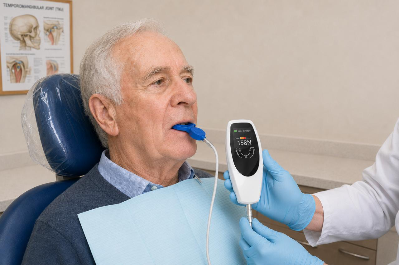
A 74-year-old patient presents for a review following implant-prosthetic rehabilitation. Absolute occlusal force measured chairside: 98 N — deep in the deficit range, where the patient can no longer properly chew ordinary food. History: 6 kg weight loss over the past year, impaired concentration, recurrent falls. The dentist noted "occlusal contacts within normal limits" and discharged the patient. And the contacts genuinely were normal. What was missing was a single number — absolute bite force — that would have revealed that behind a morphologically correct occlusion lies a serious functional deficit and a gateway to geriatric frailty, dysglycaemia, and incipient cognitive deterioration.
01DefinitionWhat exactly we measure and why it matters
Occlusal force — the maximum force generated by the masticatory muscles during intercuspation — is a measurable functional parameter, not an aesthetic indicator. The Japanese Society of Gerodontology (JSG) included it in 2016 as one of seven diagnostic criteria for oral hypofunction, a condition that precedes serious functional decline and is reversible with timely intervention (Minakuchi et al., 2018).
The crucial question, however, is what exactly you are measuring — this is what determines whether the measured number is clinically usable. Systems that capture the pressure distribution between individual teeth measure relative force: they tell you where the force goes and in what proportion, but not how much the patient actually generates. To capture occlusal force as a vital parameter you need an absolute value, expressed in newtons, provided by gnathodynamometry — the patient bites on a sensor and within a few seconds you have a reproducible number that can be shown to the patient, documented, and tracked over time.
A practical interpretive framework for absolute bite force in an adult with full dentition looks like this:
- 650–1000 N — normal bite
- 400–650 N — mild deficit: the patient begins to have difficulty chewing certain foods
- < 400 N — marked deficit with nutritional consequences
- > 1000 N — excessive force, relevant in bruxism and when planning prosthetic and implant loading
These absolute-force values must not be conflated with the thresholds of older pressure-sensitive-film methods (Dental Prescale 50H: < 200 N; Dental Prescale II: < 500 N without the pressure filter, or < 350 N with the filter) — figures from different systems are not mutually convertible (Horibe et al., 2022; Minakuchi et al., 2018).
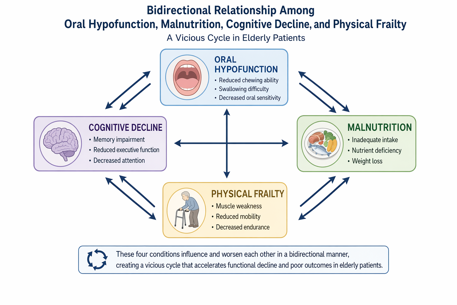
A decline in bite force is not an isolated finding — it is part of a broader syndrome encompassing reduced tongue pressure, impaired masticatory efficiency, and dysphagia. Each of these parameters individually increases the risk of systemic complications; their combination multiplies that risk.
02IndicatorsWhen to consider reduced occlusal force
Clinical indicators can be divided into four overlapping and mutually reinforcing groups.
Dental and prosthetic indicators are the most visible: extensive tooth loss without rehabilitation, worn or ill-fitting prosthetic restorations, severe attrition of occlusal surfaces, advanced periodontitis with attachment loss, or TMJ dysfunction limiting maximum intercuspation. Each of these conditions mechanically reduces the functional occlusal area and thereby the maximum achievable force.
Muscular and neurological indicators include sarcopenia of the masticatory muscles — a phenomenon that develops in older adults in parallel with generalised muscle atrophy — and neurological diseases such as Parkinson's disease, ALS, multiple sclerosis, or post-stroke motor impairment. Myopathies and myasthenia gravis are less common but are direct causes of muscular weakness.
Systemic and metabolic indicators are less intuitive for the dentist, yet fundamental: chronic malnutrition, uncontrolled type 2 diabetes mellitus, chronic kidney disease, or advanced COPD lead to generalised cachexia that also affects the masticatory muscles. Geriatric frailty is, in this context, both a cause and a consequence of oral hypofunction.
Cognitive and psychosocial indicators complete the picture: progressive cognitive deterioration, depression, or social isolation lead to neglect of dental care, avoidance of solid foods, and an overall decline in masticatory activity.
03Malnutrition and dysglycaemiaHow teeth shape metabolism
The mechanism is straightforward, but its consequences are serious. A patient with reduced occlusal force shifts to a soft, nutritionally poor diet — avoiding meat, raw vegetables, and wholegrain products. The result is a deficit in protein, fibre, B vitamins, vitamin D, and zinc. The systematic review and meta-analysis by Zelig et al. (2022) demonstrated that fully or partially edentulous older adults have a 22% higher likelihood of malnutrition or malnutrition risk compared with individuals with a functional dentition (RR 1.22; 95% CI 1.11–1.32). The overview of reviews by Kaurani et al. (2024) confirms these findings across seven systematic reviews.
The relationship with diabetes is bidirectional. Periodontitis worsens glycaemic control through a systemic inflammatory response; uncontrolled hyperglycaemia, in turn, accelerates periodontal destruction and tooth loss. The result is a closed loop in which oral hypofunction and metabolic dysregulation mutually amplify their impact.
Older adults with tooth loss are at significantly greater risk of malnutrition than individuals with a functionally adequate dentition — and this risk increases with the extent of tooth loss.
Zelig R et al. · JDR Clinical and Translational Research, 2022
04Cognition and frailtyChewing as neuroprotection
This is arguably the least intuitive yet scientifically best-supported part of the story. Mastication is not merely a mechanical process — it is a complex sensorimotor activity that, via trigeminohippocampal pathways, stimulates hippocampal neurogenesis, increases BDNF (brain-derived neurotrophic factor) expression, and maintains cerebral blood flow (Chen et al., 2015).
The meta-analysis by Cerutti-Kopplin et al. (2016), based on prospective cohort studies, demonstrated that individuals with fewer than 20 teeth have a 26% higher risk of cognitive decline (HR 1.26; 95% CI 1.14–1.40) and a 22% higher risk of dementia (HR 1.22; 95% CI 1.04–1.43) compared with individuals with ≥ 20 teeth. The dose-response meta-analysis by Qi et al. (2021) further showed that each tooth lost increases the relative risk of cognitive decline by 1.4% — and edentulous patients face a 1.54-fold higher risk of cognitive impairment.
The key finding is that prosthetic rehabilitation mitigates this risk: patients with dentures show a markedly lower risk of cognitive decline than edentulous individuals without any prosthetic replacement (Qi et al., 2021). This repositions prosthetic treatment from the category of comfort to that of preventive medicine — and makes restoring occlusal force a measurable, demonstrable treatment goal.
Oral frailty — defined in Tanaka's framework as concurrent deterioration in ≥ 3 of six oral-function parameters (number of teeth, chewing ability, articulatory motor function, tongue pressure, subjective difficulty with eating, and difficulty swallowing) — is a strong predictor of physical frailty and mortality. It is a construct distinct from JSG oral hypofunction (7 criteria), even though the two domains overlap. Tanaka et al. (2018) in the longitudinal Kashiwa study (n = 2,011) demonstrated that oral frailty is associated with a 2.4-fold higher risk of physical frailty, a 2.2-fold higher risk of sarcopenia, and a 2.2-fold higher risk of mortality.
05Clinical recommendationsWhat this means for everyday practice
- Measure absolute occlusal force routinely — in newtons, chairside, at the initial examination and after prosthetic rehabilitation. In patients over 65, pay particular attention to sudden drops in force over time. A reproducible number the patient sees on the display is also the strongest argument for accepting treatment: a deficit that can be shown is treated more willingly than one the patient merely hears about.
- Screen for oral hypofunction according to JSG criteria (Minakuchi et al., 2018): combine assessment of occlusal force with tongue pressure, masticatory efficiency, and dysphagia screening.
- A sudden drop in occlusal force without an obvious dental cause is a red flag for systemic disease — consider referral to a general practitioner or geriatrician.
- Prosthetic rehabilitation is neuroprotection: do not wait for "ideal conditions" — even partial functional rehabilitation reduces the risk of cognitive decline and malnutrition. With absolute gnathodynamometry, the benefit is also demonstrable as a concrete increase in force in newtons before and after treatment.
- Interdisciplinary collaboration with a nutritionist, geriatrician, or neurologist is the standard of care — not the exception — for patients with oral hypofunction.
- Document functional parameters, not just morphology: occlusal force, tongue pressure, and masticatory scores are clinically relevant data that belong in the patient record alongside radiographic findings.
- Educate patients: explain that dental care and prosthetic rehabilitation have a direct impact on overall health, cognitive function, and length of active life. The number on the display makes this connection tangible.
06References
-
Minakuchi S et al. Oral hypofunction in the older population: Position paper of the Japanese Society of Gerodontology in 2016. Gerodontology. 2018;35(4):317–324. doi:10.1111/ger.12347
-
Horibe Y, Matsuo K, Ikebe K, Minakuchi S, Sato Y, Sakurai K, Ueda T. Relationship between two pressure-sensitive films for testing reduced occlusal force in diagnostic criteria for oral hypofunction. Gerodontology. 2022;39(1):3–9. PMID: 33555057. doi:10.1111/ger.12538
-
Zelig R et al. Tooth Loss and Nutritional Status in Older Adults: A Systematic Review and Meta-analysis. JDR Clinical and Translational Research. 2022;7(1):4–15. doi:10.1177/2380084420981016
-
Kaurani P et al. Association of tooth loss and nutritional status in adults: an overview of systematic reviews. BMC Oral Health. 2024;24:897. doi:10.1186/s12903-024-04602-1
-
Cerutti-Kopplin D et al. Tooth Loss Increases the Risk of Diminished Cognitive Function: A Systematic Review and Meta-analysis. JDR Clinical and Translational Research. 2016;1(1):10–19. doi:10.1177/2380084416633102
-
Qi X et al. Dose-Response Meta-Analysis on Tooth Loss With the Risk of Cognitive Impairment and Dementia. Journal of the American Medical Directors Association. 2021;22(10):2039–2045. doi:10.1016/j.jamda.2021.05.009
-
Tanaka T et al. Oral Frailty as a Risk Factor for Physical Frailty and Mortality in Community-Dwelling Elderly. The Journals of Gerontology: Series A. 2018;73(12):1661–1667. doi:10.1093/gerona/glx225
-
Chen H et al. Chewing Maintains Hippocampus-Dependent Cognitive Function. International Journal of Medical Sciences. 2015;12(6):502–509. doi:10.7150/ijms.11911
-
Dibello V et al. Oral frailty indicators to target major adverse health-related outcomes in older age: a systematic review. Geroscience. 2023;45(2):663–706. doi:10.1007/s11357-022-00663-8
-
Nakamura T et al. Oral dysfunctions and cognitive impairment/dementia. Journal of Neuroscience Research. 2021;99(2):518–528. doi:10.1002/jnr.24745

🌍 Cicero System🎓 Dental Education | 🛒 Procurement | 🏥 Clinic Management