
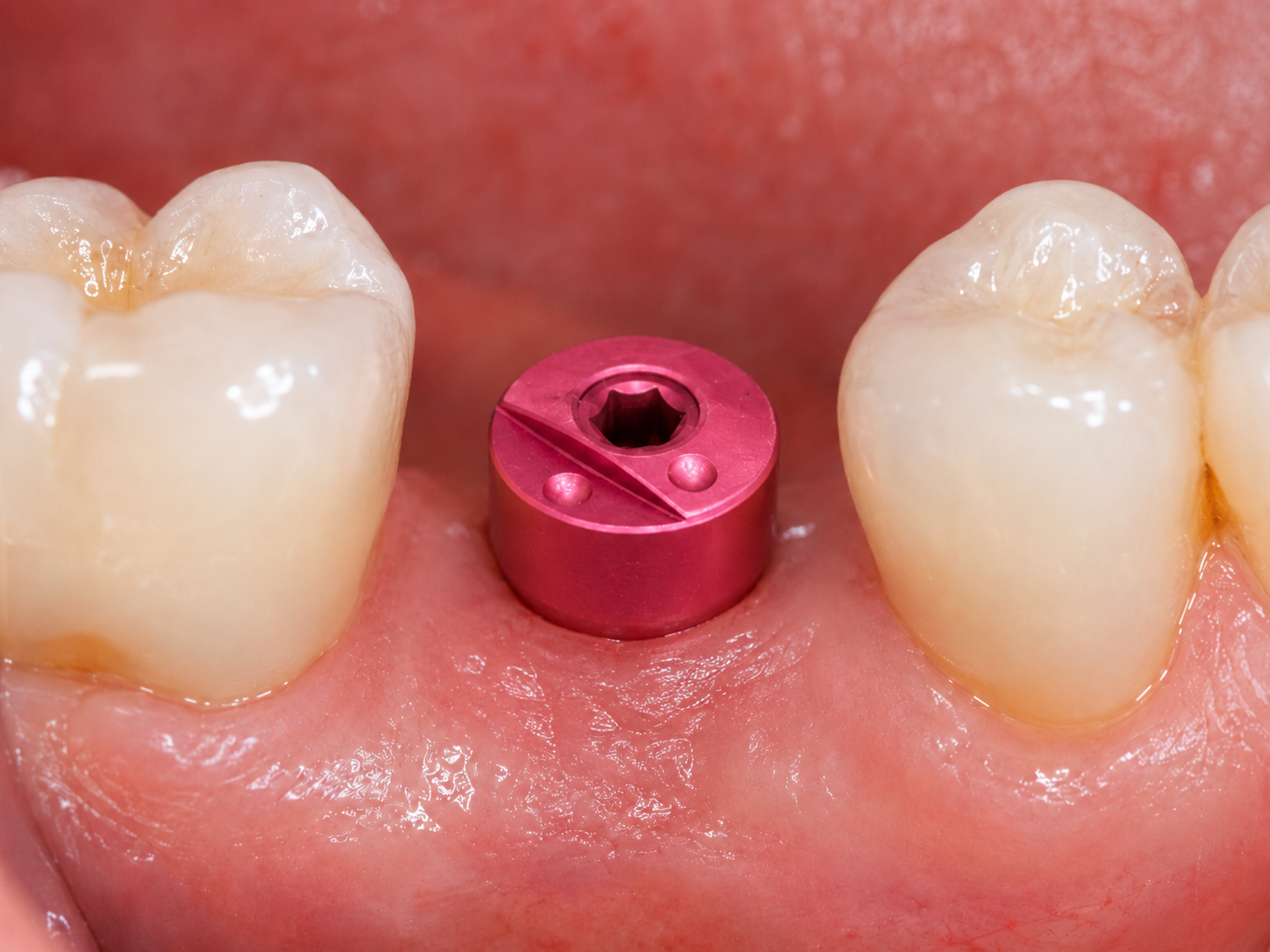
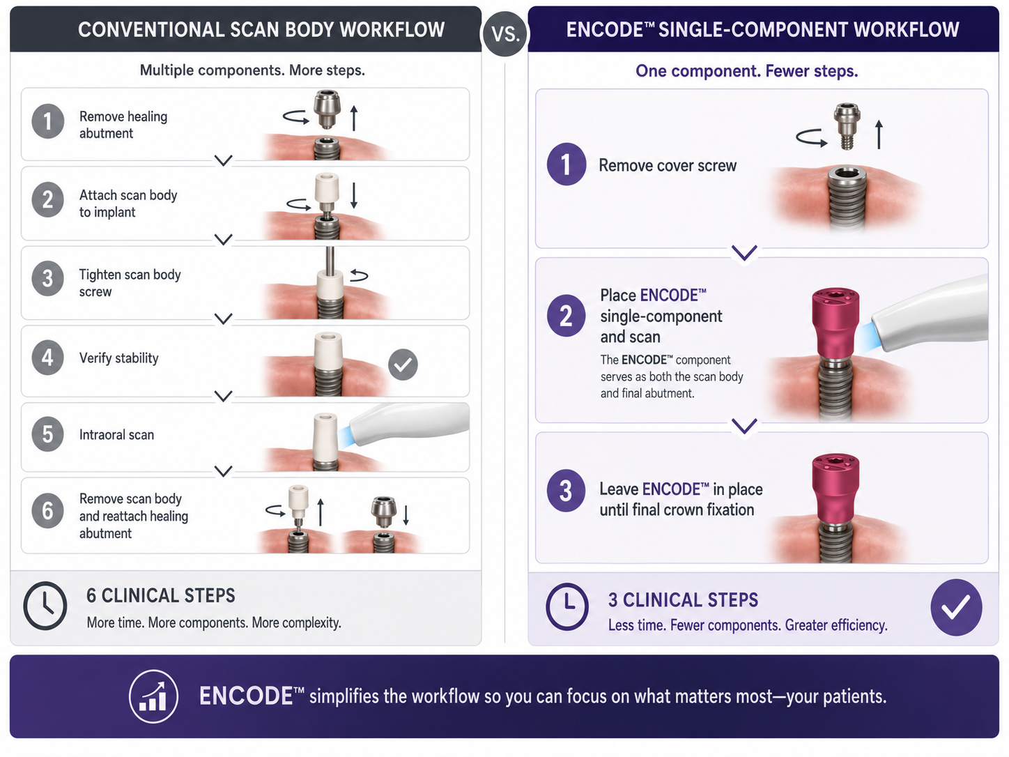
Walk into most implant workflows and the sequence is predictable: place a healing abutment, let the tissue mature, remove the healing abutment, connect a scan body, scan, remove the scan body, reconnect the healing abutment. Repeat for every appointment. The Encode system from ZimVie — formerly Biomet 3i — quietly eliminates several of those steps, and the mechanism behind it is worth understanding properly.
01The Classic ApproachHow a Conventional Scan Body Works
A conventional scan body (also called a scanbody or ISB — implant scan body) is a precision-machined component that connects directly to the implant or to a multi-unit abutment. Its geometry is unique and asymmetric: the intraoral scanner reads the shape, cross-references it against the manufacturer's digital library, and calculates the exact three-dimensional position and angulation of the implant platform in space.
The system works well. Systematic review evidence confirms that intraoral scanning with dedicated scan bodies delivers clinically acceptable accuracy for single-implant and short-span cases. The limitation is procedural: the healing abutment must come off, the scan body goes on, the scan is taken, and then the healing abutment is replaced. Every component swap carries a small risk of disturbing the peri-implant tissue cuff, and in a busy clinical day those extra steps accumulate.
02The Encode ConceptOne Component, Two Functions
The Encode Healing Abutment does something architecturally different. Its coronal surface is machined with a proprietary pattern of concentric rings and geometric features — the "code" — that encodes the implant's platform size, connection type, and rotational orientation directly into the abutment's topography. When you scan the patient with an intraoral scanner, the scanner reads that coded surface exactly as it would read a conventional scan body.
The result: the healing abutment never needs to be removed for the impression. The patient heals around a component that is simultaneously shaping the emergence profile and acting as the reference geometry for the digital workflow. One appointment step disappears entirely.
ZimVie's digital library maps each Encode abutment variant to the corresponding implant data, so the CAD software receives the same positional and angular information it would get from a dedicated scan body — platform diameter, implant axis, rotational index — without any component swap.
03What Changes ClinicallyThe Practical Difference
The most immediate benefit is tissue stability. Every time a healing abutment is removed and replaced, there is a risk of micro-trauma to the forming sulcular epithelium and connective tissue cuff. Research on scan body design confirms that the number of component exchanges is a procedural variable worth controlling — particularly in the aesthetic zone where the emergence profile is critical.
Beyond tissue management, the Encode system reduces chairtime and the risk of component mix-up. In a multi-implant case, keeping track of which scan body belongs to which implant position is a real source of error. With Encode, the coded surface is already in place and already indexed to that specific implant — there is nothing to swap, nothing to lose, and no chance of connecting the wrong component.
The system is also compatible with ZimVie's full digital workflow: scans feed directly into design software with the implant library pre-loaded, and the restorative team receives an accurate virtual model without a physical impression or a stone cast.
04Scan Body Design MattersWhat the Evidence Says
Recent systematic review evidence makes clear that scan body geometry — height, diameter, surface features, and library congruence — significantly affects the accuracy of digital implant positioning. The Encode surface is specifically engineered to be readable by current intraoral scanners, with enough geometric contrast to allow reliable identification even when the abutment is partially surrounded by soft tissue.
For single implants and short-span cases, the accuracy of a well-designed encoded healing abutment is clinically comparable to a dedicated scan body. For full-arch cases, the same principles that apply to conventional scan bodies apply here: scanning strategy, operator experience, and scanner model all influence the final result.
Scan body design — including geometry, material, and library match — is a primary determinant of digital implant impression accuracy.
Mizumoto R.M. · Journal of Prosthetic Dentistry, 2018
05Availability and CompatibilityWho Is It For
The Encode system is designed for ZimVie's Certain® and Tapered Screw-Vent® implant lines. It is available in a range of platform diameters and emergence profile heights to accommodate different tissue depths and aesthetic requirements. The system requires that the treating clinician's CAD/CAM software or lab partner has access to the ZimVie digital library — a standard requirement for any manufacturer-specific scan body workflow.
For clinicians already working within the ZimVie ecosystem, Encode is a straightforward upgrade to the restorative protocol. For those evaluating implant systems, the integrated healing-and-scanning function is a meaningful differentiator — particularly in practices that prioritize digital efficiency and tissue-friendly workflows.
The core idea is simple but consequential: instead of adding a component to take the impression, you design the healing component so the impression is already built in.

👨⚕️ doc. MUDr. Radek Mounajjed DDS., PhD. 🦷 D.C.M. Clinic 🎓 Associate Professor, Palacký University Olomouc, Czech Republic 📚 CICERO Cofounder ⚖️ Certified Court Expert in Dentistry
View educator profile